
Researchers Pioneer 3D Human Liver Models to Improve Drug Safety Testing
A research team at the FAMU-FSU College of Engineering has moved closer to transforming pharmaceutical drug safety testing with a new 3D liver cell model that predicts drug-induced liver toxicity more accurately than current methods.
The study, led by Jamel Ali, director of a new center for pharmaceutical engineering and an associate professor
in the college’s Department of Chemical and Biomedical Engineering, was recently published in ACS Pharmacology & Translational Science.
“Our work involves innovative biomaterials that brings us closer to functional tissues
that could transform patient care,” Ali said. “There are hurdles ahead, but each breakthrough
fuels our hope for the future of medicine. We want to build tissues that not only
mimic the real human liver, but could one day save lives.”
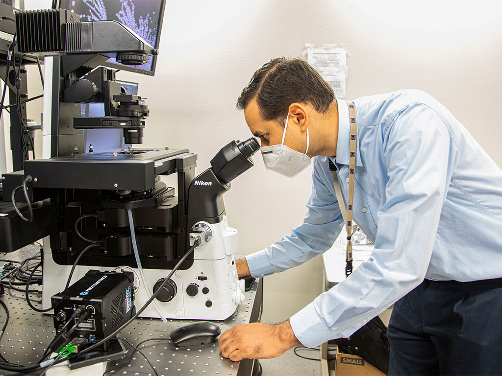
 Jamel Ali, is a FAMU professor in the FAMU-FSU College of Engineering. (Photo courtesy
of FAMU-FSU College of Engineering)
Jamel Ali, is a FAMU professor in the FAMU-FSU College of Engineering. (Photo courtesy
of FAMU-FSU College of Engineering)
Why Current Drug Safety Testing Falls Short
The liver is the body’s primary organ for filtering toxins, but even medicines designed to help can cause unexpected and dangerous liver damage—a condition called hepatotoxicity. For decades, researchers have relied on flat, two-dimensional cell layers or animal models to screen new drugs before they reach patients.
Those methods sometimes miss subtle warning signs, the researchers say, putting patients
at risk when drugs reach the market. The limitations carry significant cost implications:
the average expense of developing and gaining FDA approval for a new drug ranges from
approximately $1 billion to more than $2 billion, according to estimates from multiple
independent analyses, with failed or withdrawn drugs adding to that figure.
A Tallahassee Lab’s 3D Approach to Hepatotoxicity Screening
The research group’s method uses 3D clusters of liver cells, known as spheroids, grown within a soft, gelatin-based biomaterial. This scaffold better simulates the structural environment of the human body, allowing liver cells to form small clusters that behave much more like actual liver tissue than cells grown flat on a dish.
The 3D liver cell culture the team developed keeps human liver cells viable and functional for close to a month—considerably longer than standard lab methods. Critically, its toxicity results align closely with what has been documented in real human patients.
When the team tested the system against five drugs known to harm the liver, the 3D culture identified toxic doses that tracked closely with real human treatment levels. By comparison, standard 2D cell cultures tended to make those same drugs appear more dangerous than they actually are—a distortion that can skew development decisions early in the pipeline.
In an additional test, when human liver tissues grown with this method were implanted in mice, they survived, developed new blood vessels, and were not rejected by the immune system. That result suggests the approach could support drug testing in living animals using human tissue, bridging a key gap between lab and clinical outcomes.

Graduate Researchers Drive the Work Forward
Mary Jean Savitsky, a fourth-year Ph.D. student in biomedical engineering at Florida State University (FSU) who works with Ali’s group, described the project’s scope.
“This project, focused on developing a bioink for 3D printing healthy human liver tissue, marks a significant advance toward the future,” Savitsky said. “By enabling the creation of functional human tissue outside the body, our work helps establish better liver models, reduces reliance on animal testing, and furthers the broader goal of tissue regeneration and personalized medicine.”
Beyond Hepatotoxicity: Potential Applications Across Organ Systems
The 3D liver model has implications well beyond liver toxicity screening, according to Ali.
“While this system is currently focused on liver tissue and developing improved in vitro models for hepatotoxicity drug screening and in vivo liver function restoration, our approach has broader potential,” Ali said. “In earlier work, similar tissue engineering strategies helped us replicate pancreatic functions and reverse hyperglycemia in mice with Type 1 diabetes. Now, we’re working to adapt these models to test other organs and cancer tissues.”
A Platform Compatible With Standard Lab Equipment
Navneet Kaur, the study’s lead author and a postdoctoral scholar at Penn State University, worked with Ali and Savitsky during her time as a researcher at the National High Magnetic Field Laboratory in Tallahassee. She says the platform is ready for use in working labs today.
“This platform is compatible with standard lab equipment, allowing high-throughput testing of many drug compounds at once,” Kaur said. “It’s more cost-effective than previous 3D materials and demonstrated consistent results, so we anticipate these models will soon become a standard tool in drug development.”
That readiness for real-world use sets this work apart from earlier 3D systems, Kaur explained.
“Traditional 2D cell models are easy to use and widely established, but they don’t capture the three-dimensional complexity of the human body,” she said. “Our 3D models provide a much clearer window into how drugs will behave in real patients and whether they may be toxic.”
Challenges Remain on the Path to Clinical Use
Despite the results, Ali is direct about what the team has yet to prove.
“Although these model systems represent a substantial step forward, there are still significant challenges ahead. It remains uncertain whether the complexities of human liver function can be fully replicated or if these systems will ultimately prove clinically effective for restoring liver function in patients,” he said. “However, our ongoing research with human liver and other tissues, together with our innovative biomaterials, aims to create functional tissues that could one day be used in clinical practice.”
Multi-Institutional Collaboration
The study is the product of a team spanning multiple institutions. Co-authors include Navneet Kaur (Penn State University), Mary Jean Savitsky and Jamel Ali from the FAMU-FSU College of Engineering, Michael Lipscomb from the University of Minnesota, and Dazhi Yang from Acrogenic Technologies Inc. Support for this research was provided by the National Institutes of Health, the NSF FAMU CREST Center, the National High Magnetic Field Laboratory, the Grainger Foundation and NASA.
Editor’s Note: This article was edited with a custom prompt for Claude Sonnet 4.5,
an AI assistant created by Anthropic. The AI optimized the article for SEO discoverability,
improved clarity, structure and readability while preserving the original reporting
and factual content. All information and viewpoints remain those of the author and
publication. This article was edited and fact-checked by college staff before being
published. This disclosure is part of our commitment to transparency in our editorial
process.